
For years, orthopaedic doctors at a government hospital in Bengaluru had noticed that about 11% of their daily patients comprised people who had developed a debilitating bone deformity after going to quacks to treat fractures.
In such cases, the only treatment was conventional, inexact surgeries that did not guarantee total success.
Now, working with scientists, doctors at the Sanjay Gandhi Institute of Trauma and Orthopaedics (SGITO) have fashioned a new way to fix this problem.
A three-year clinical study, which began in September, has so far treated five patients sourcing tools from three places: Indian Institute of Science (IISc), industries in Peenya, Bengaluru, and software from Belgium.
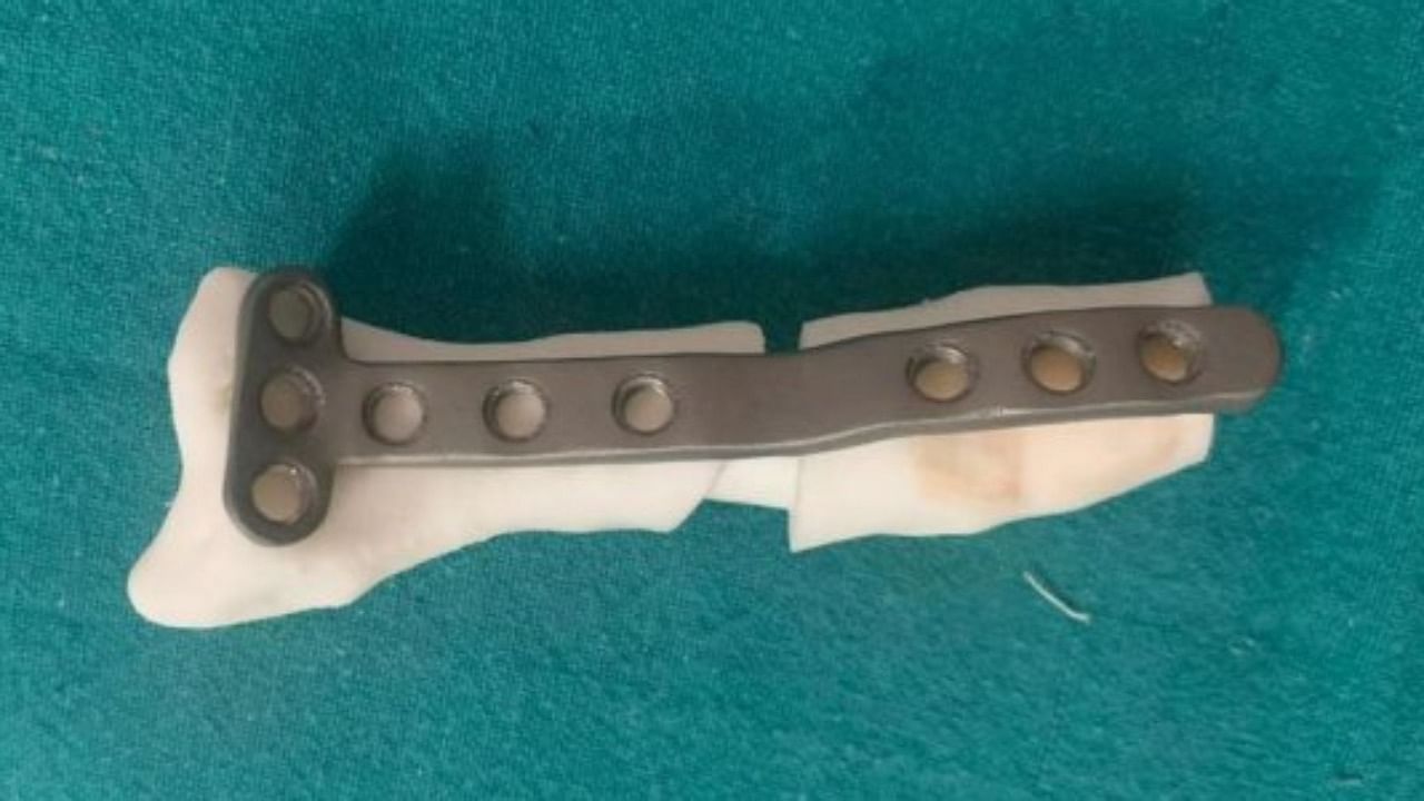
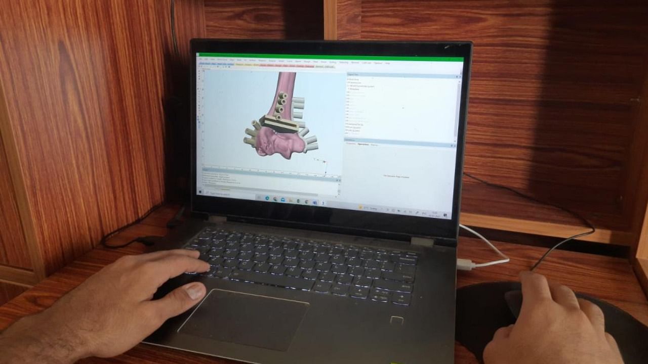
Advanced 3D printing technology from the IISc is used to create a mold of the fractured bone and fabricate surgery guides. Next, industries in Peenya create a metal implant to correct the deformity with the help of a 3D manipulation software from Belgium, which allows researchers to “correct” the deformed bone in virtual space.
Dr Sathya Vamsi Krishna from the SGITO and an authority on upper limbs said that four to five of 35 OPD patients that he sees per day on average were individuals with complex bone deformities. “In India, about 30-40% of people go to quacks for fracture treatment,” he added.
“If fractures heal in awkward positions, this creates a deformity,” said Dr H S Chandrasekhar, a leading orthopaedic surgeon and Director of SGITO.
This was the case with 16-year-old Malina (name changed) who suffered a small fracture to her right wrist in July.
Within days, a dull pain became searing whenever she tried to move her hand. “She became unable to use her right hand and could no longer write in class,” her father, Mansoor said, adding that he delayed treatment for two months believing the pain would go away.
By September, Malina’s fractured wrist bone had grown at an awkward angle that required urgent surgery. She became patient no. 3 in the new clinical trial.
“While a CT-Scan can give a three-dimensional picture of the deformed bone, correcting this in surgery is difficult,” Dr Krisha said.
He added that he only realised that 3D printing could help after stumbling upon a paper about 3D-printed bone plates by IISc scientists.
Associate Professor Kaushik Chatterjee of IISc’s Department of Materials Engineering and Center for BioSystems Science and Engineering, who was corresponding author for that paper, explained how the new method works.
The fracture is scanned and the results uploaded to the software for analysis in a three-dimensional space.
Once the doctors have decided on the best way to correct the defect using a metal brace implant, the custom specifications of the deformed bone and implant are sent to IISc and the institute’s industry partner for fabrication.
“The critical step is the planning stage. The surgery itself may take an hour or less, but planning out the surgery, using these models and designing the metal implants to support the bone takes about two weeks,” Dr Krishna said.
Two key students at IISc, Saurabh Gupta and Navaneeth Holla, design the custom implants.